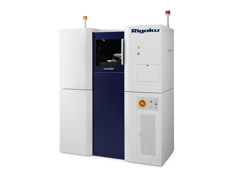
True submicron CT scanner with contrast-enhancing X-ray anode
3D X-ray microscope
Rigaku nano3DX is a true submicron resolution CT (computed tomography) scanner. The parallel beam geometry combined with an ultrabright 1200 W rotating anode X-ray source enhances the contrast of soft materials, which are normally difficult to image using high-energy X-ray sources. The Cu (8 keV) X-ray anode covers a wide range of soft materials, combined with one of these materials—Cr (5.4 keV), Mo (17 keV) or W—to image ultralow or medium density samples. With the highest magnification lens, the nano3DX can achieve 188 nm voxel resolution and true submicron (500 nm) spatial resolution.
nano3DX Overview
How do I achieve high resolution?
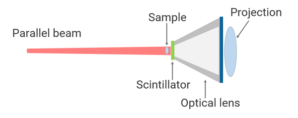
Rigaku nano3DX uses parallel beam geometry. This geometry uses an optical lens to magnify the sample image. It does not use the X-ray beam divergence and eliminates blurring caused by the X-ray focus size and drift. With the 20X magnification lens, you can achieve 188 nm voxel resolution and true submicron (500 nm) spatial resolution.
At this resolution, you can see individual carbon fibers (~7.5 microns), the intricate structures of seeds, small insects, etc.
How do I change X-ray anodes?
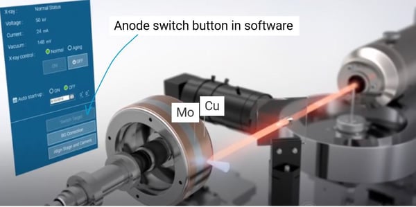
Rigaku nano3DX is equipped with a dual-wavelength rotating anode X-ray generator, MicroMax-007 HF. The rotating X-ray anode is made of Cu, providing Cu characteristic radiation of 8 kV, combined with a second anode material, Cr 5.4 kV, Mo 17 kV, or W for Bremsstrahlung radiation for wider energy coverage. You can switch between two radiations with a simple click on the instrument control software.
You can image an even 5% density difference in organic materials by using optimized radiation and differentiate amorphous and crystalline phases, high- and low-density polyethylene, etc.
Do I need to prepare my sample in a special way?
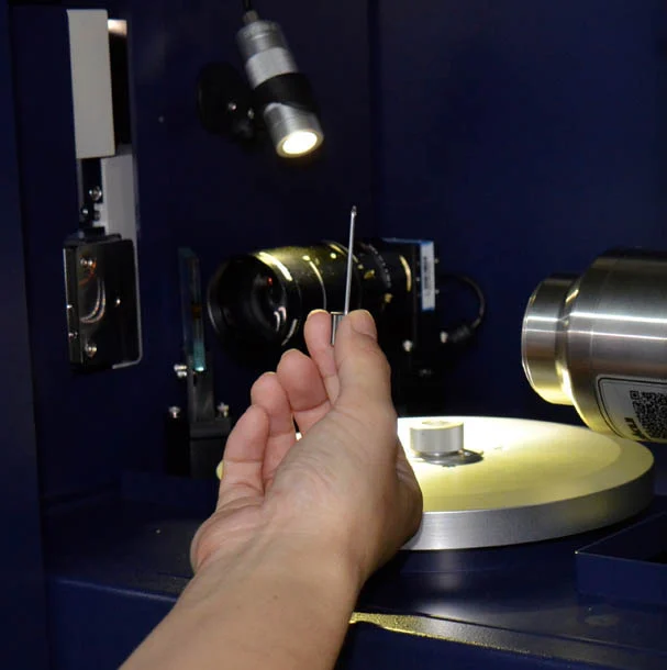
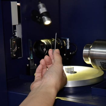
No, X-ray CT measurements require minimum to no sample preparation. As far as the sample size is close to the FOV (field of view), you can mount the sample on the sample stage with a small pin as is, and you are ready to run a CT scan.
Because X-ray CT experiments are done in the air and do not require conductivity on the sample as is the case for SEM (scanning electron microscopy), organic samples such as plants can be measured without a drying or coating process.
nano3DX Features
- 188 nm voxel / 500 nm spatial resolution
- Parallel beam geometry for true submicron resolution
- Best X-ray energy for low-density materials
- Selectable pseudo-monochromatic X-ray radiations
- Shorter scan time
- Ultrabright 1200 W X-ray source and sCMOS to increase the X-ray count and detection efficiency
nano3DX Specifications
| Voxel resolution | 188 nm — 12 μm | |
|---|---|---|
| Field of view (FOV) | 0.9 — 14.4 mm | |
| Field of view (FOV) 0.66 — 20 mm Maximum sample size | 20 mm diameter x 40 mm height | |
| Geometry | Parallel beam geometry | |
| X-ray source | 1200 W rotating anode microsource | |
| X-ray energy | Primary source: Cu (8 kV) Second source: Choose one from Cr (5.4 kV), Mo (17 kV), and W (Bremsstrahlung radiation with applied voltage 60 kV) |
|
| Lenses | 1.25X, 2.5X, 5X, 10X, 20X | |
| Detector | High-resolution sCMOS | |
| Detector pixel size | 3.76 µm | |
| Detector size | 4800 x 4000 pixels | |
| Dimensions | 1300 (W) x 1880 (H) x 655(D) mm (PC, chiller, vacuum pump not included) | |
| Weight | Approx. 600 kg | |